Anfatec Logo

Confocal Scanning Imaging

Click to enlarge Click to enlarge

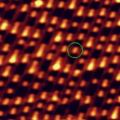
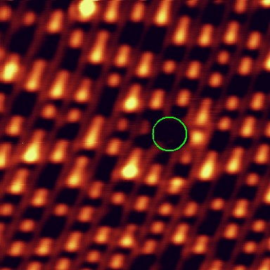
Confocal Imaging of a CD-ROM

The images on the left show confocal scans of the bit patters of a commercial CD-ROM. A green laser (532 nm) was focussed via the bottom objective on the CD sample. The back reflection is guided via the beam splitter and a periscope to a port with a photo detector.

The images represent a sample size of 50 µm x 50 µm and 20 µm x 20 µm respectively.

Click to enlarge Click to enlarge

Writing on a CD-R

After a confocal scan (left image), the sample was treated with an intense laser beam. The change of the bit patern is highlighted by a green circle.

The image size is 20 µm x 20 µm.

Click to enlarge

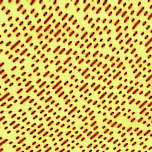
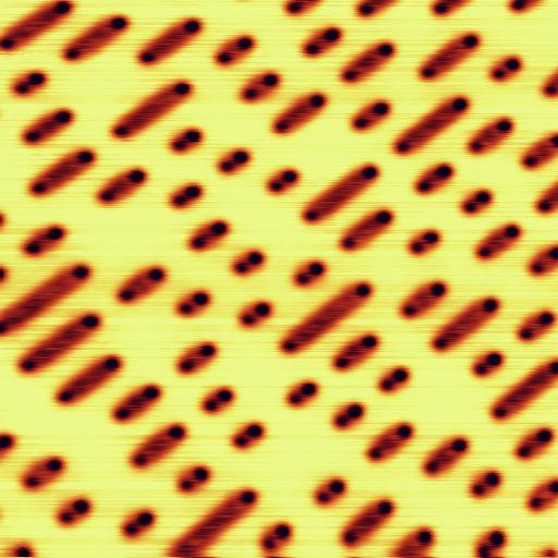
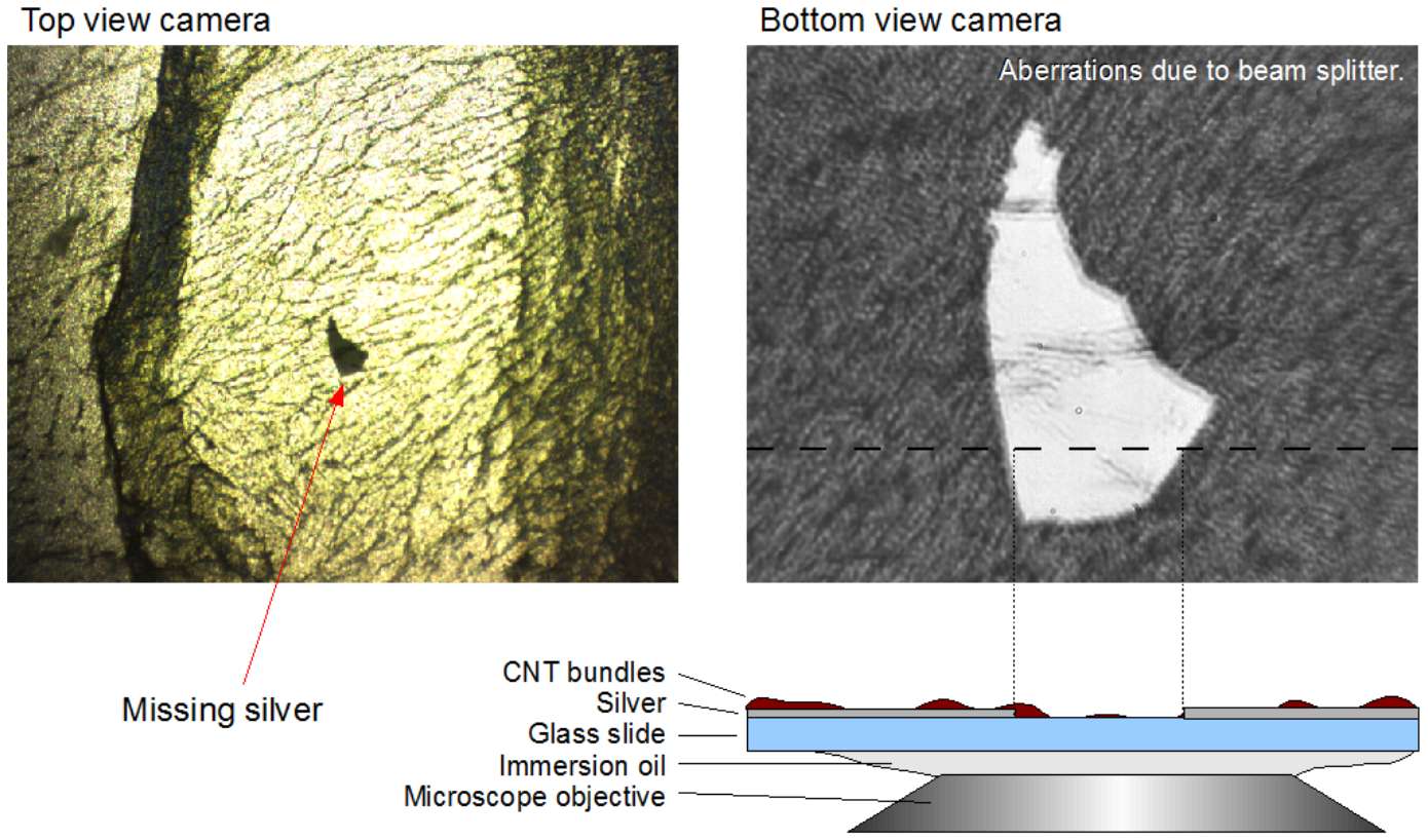
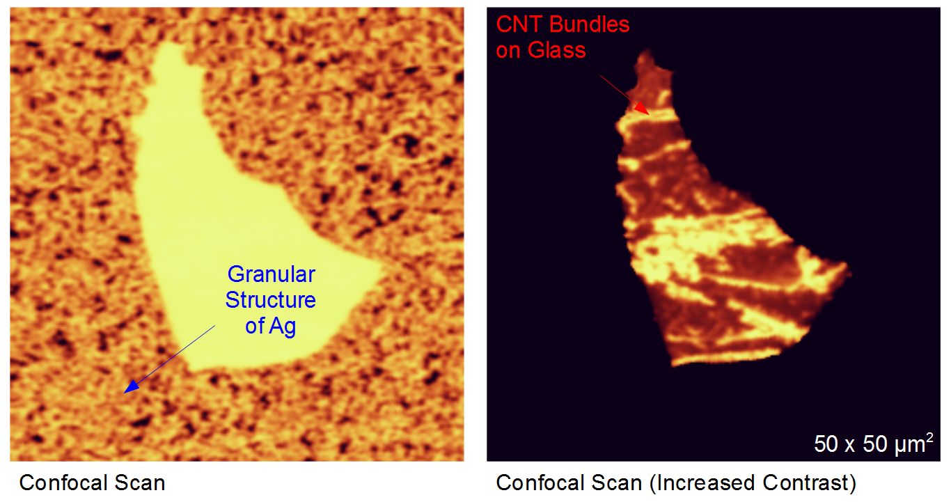
Confocal Scan on Carbon Nanotube (CNT) Bundles on a Glass/Silver Substrate

The images on the left are obtained by the top (left) and the bottom (right) objective of the VistaScope . Additionally a sketch of the sample layer structure is shown below the bottom view image.

Click to enlarge

The confocal scan on the left shows the inverted image of silver-free part of the sample. Here, the glass is covered by CNT bundles only. The silver layer gives rise to a high back reflection intensity of the confocal laser. Due to the inversion of the intensity signal, the glass part of the sample is very bright. The granular structure of the silver layer is nicely resolved. After increasing the image contrast, the CNT bundles are visible as well. At this stage, imaging of a confocal Raman spectrum is possible (see also Applications > Raman Mapping).

The image size is 50 µm x 50 µm.

Overview Applications Technical Details